Sachee Bhanu Piyasiri1, Thisum Nilakshi Samaranayake1*, Hermali Silva1, Nuwani Harshamali Manamperi2, Nadira Darshani Karunaweera1
1 Faculty of Medicine, Department of Parasitology, University of Colombo, Colombo, Sri Lanka
2 Faculty of Medicine, Department of Parasitology, University of Kelaniya, Ragama, Sri Lanka
Reference: Piyasiri SB, Samaranayake TN, Silva H, Manamperi NH, Karunaweera ND. ELISA‐based evaluation of antibody response to Leishmania in a region endemic for cutaneous leishmaniasis. Parasite Immunology. 2022 Sep;44(9):e12940.
https://pubmed.ncbi.nlm.nih.gov/35836368/
Summary
This research aimed to evaluate anti-leishmanial IgG antibody responses as a biomarker for active cutaneous leishmaniasis (CL) and as a measure of exposure to Leishmania. The study involved analyzing sera from 50 untreated CL patients, 140 patients under treatment, and 280 healthy individuals in endemic regions using an in-house ELISA. The assay demonstrated high diagnostic performance in identifying exposure in endemic individuals (sensitivity: 98%, specificity: 90.3%). Anti-IgG antibody levels were higher and persisted longer in untreated patients compared to those under treatment. In patients under treatment, antibody levels negatively correlated with the total treatment duration. The study concluded that the anti-leishmanial IgG response in L. donovani-induced CL is transient and unlikely to confer protective immunity. The optimized serological assay may be useful for diagnosis and monitoring treatment response in CL in endemic settings.
The study population included CL patients and healthy individuals in Sri Lanka, an endemic region for CL caused by L. donovani. The in-house ELISA assay showed promise in distinguishing active infection from exposure or asymptomatic infections. The research provides insights into the dynamics of anti-leishmanial IgG antibodies during different stages of CL and suggests a potential role for the assay in diagnosing and monitoring CL in endemic settings. Further validation and antigen profiling are recommended for standardizing the assay and exploring its application in varied endemic settings.
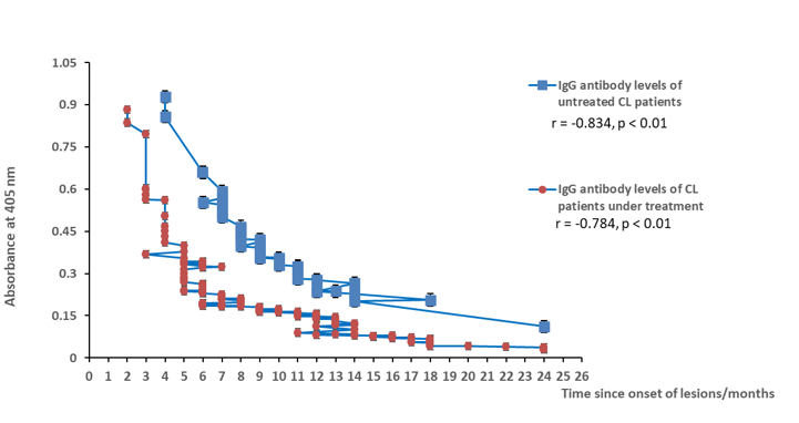
Figure: Clinical correlations of CL patients with levels of serum anti- leishmanial IgG of untreated CL patients (Pearson’s r= −0.834, p ˂ 0.01) and patients under treatment (Pearson’s r = −0.784, p ˂ 0.01) with the lesion duration/months.
